Retroperitoneoscopic ureterolithotomy (RPU) was performed in 30 patients (20 men, 10 women). The indications for this operation were large (more than 1.5 cm) stones of the upper and middle third of the ureter that had grown into the mucous membrane and were not amenable to extracorporeal shock wave lithotripsy or endoscopic removal. In all cases, due to the large size, high density and long standing time of the stones, RPU was originally planned. The size of the stones was from 15 to 32 mm, in 18 cases they were localized in the upper third, in 12 cases in the middle third of the ureter. The results of RPU were compared with the results of a retrospective group of 26 patients who underwent open surgery. The stones were the same size. The duration of the operation, the incidence of complications, the dose of analgesics used, the duration of the postoperative stay in the hospital and the rehabilitation of patients were compared. RPU was successful in 28 patients (93.3 %). In 2 cases, the stones were not found due to their movement into the pelvis. These stones were subsequently crushed against the background of the stent. In the postoperative period, urine leakage was noted in 2 patients, which stopped after PC nephrostomy. In the groups of RPU and open ureterolithotomy, there were no significant differences in the duration of the operation, the incidence of complications, the dose of analgesics used, the terms of postoperative stay in the hospital and the rehabilitation of patients. RPU is an effective, safe method for the treatment of large, ingrown ureteral stones and may become an alternative method of treatment in the hands of urologists who are proficient in laparoscopic surgical techniques.
Keywords: retroperitoneoscopic ureterolithotomy (RPU), extracorporeal shock wave lithotripsy, ureteral JJ stent, lithotripsy
Introduction. Currently, the main methods of treating patients with ureterolithiasis are remote (extracorporeal) and contact ureterolithotripsy. However, in a certain group of patients with large (more than 1.5–2.0 cm) ureteral stones that are in one place for a long time, these surgical interventions are not always effective. In such cases, laparoscopic ureterolithotomy is an alternative treatment [1–4]. In surgery of the abdominal cavity and retroperitoneal space, laparoscopic operations are traditionally performed by transabdominal access. However, M. Bartel in 1969 proposed a retroperitoneal approach [5]. The first reports of performing lumboscopic ureterolithotomy appeared in 1979. [6]. Due to the possibility of urine leakage from the ureteral incision after ureterolithotomy, the retroperitoneal approach is more preferable.
However, some urologists suggest performing laparoscopic ureterolithotomy through the transperitoneal approach [7, 8]. In their opinion, the frequency of postoperative complications and the results of surgical interventions are comparable with those after retroperitoneal stone removal. The advantages of the lumboscopic method compared to traditional open ureterolithotomy are to reduce the surgical wound and trauma during tissue mobilization, and reduce the duration of the operation.
Material and methods
From 2010 to 2013, retroperitoneoscopic ureterolithotomy was performed in 30 patients (20 men, 10 women) at the Republican Specialized Center of Urology. Indications for this operation were dense impacted stones of the proximal ureter of large sizes. Due to the characteristics of the stones (dimensions greater than 15 mm, high density, duration of standing for more than 4 months), patients were initially planned for laparoscopic ureterolithotomy. Stone sizes ranged from 15 to 32 mm (average 23 mm). In 18 cases, they were localized in the upper third of the ureter, and in 12 cases, in the middle one, above the intersection of the ureter with the iliac vessels. All patients had calculi that were X-ray positive. Operations were performed only by retroperitoneal access in the position of the patient on his side (Fig. 1).
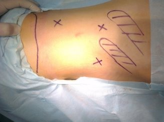
Fig. 1: X - the proposed points for the installation of trocars
The first 10 mm trocar was placed under the rib along l. axillaris posterior. Using a laparoscope with constant air insufflation up to 12 mm Hg. a working cavity was created in the retroperitoneal space. Then two more trocars 5 and 10 mm for working instruments were installed (Fig. 2).
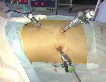
Fig. 2: Trocars are placed in the lumbar region
The peritoneum was exposed and retracted medially. The ureter was identified in the retroperitoneal fatty tissue, it was isolated until the stone was found, which was removed through one of the laparoscopic ports (Fig. 3).
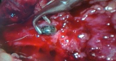
Fig. 3: The ureter is exposed, ureterolithotomy and removal of the stone with a forceps
The ureteral defect was closed with a continuous suture (Vicryl 4/0). The operation ended with drainage of the retroperitoneal space. The results of RPU were compared with the results of a retrospective group of 26 patients who previously underwent open surgery in our clinic before the introduction of modern methods of treating ureterolithiasis. Their stone sizes ranged from 14 to 31 mm (average 22 mm). All surgical interventions were performed under general anesthesia by urologists trained in foreign clinics. A comparative analysis was carried out of such parameters as the duration of the operation, the frequency of complications, the doses of analgesics used, the terms of postoperative stay in the hospital and the patient's rehabilitation. To compare the obtained quantitative data after retroperitoneoscopic and open ureterolithotomy, the Student's test was used. At p<0.05, the studied indicator was significant.
Results
The results of laparoscopic ureterolithotomy were successful in 28 (93.3 %) patients. In two cases, the stone was relocated proximally into the pelvicalyceal system, in connection with which a ureteral JJ stent was placed and, subsequently, the stone was subjected to extracorporeal shock wave lithotripsy. In the postoperative period, 2 (6.6 %) patients had urine leakage, which stopped after the installation of a PC nephrostomy. We present a clinical case of successful removal of a stone in the upper third of the ureter by lumboscopic access. Patient X., aged 56, was hospitalized in the RSCU in a planned manner with pain in the lumbar region on the right. From the anamnesis, for the first time, renal colic on the right was noted 6 months ago, symptomatic therapy was carried out at the urologist at the place of residence, but no stone discharge was noted. Due to the cessation of the pain syndrome, further examination and treatment was not carried out. Examination at the RSCU revealed a large stone in the projection of the upper third of the right ureter on a plain radiograph (Fig. 4). On the urograms, the function of the left kidney is satisfactory, on the right, contrasting is slow (Fig. 5).
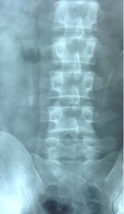
Fig. 4: The shadow of the calculus in the projection of the upper third of the ureter
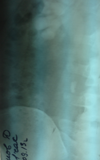
Fig. 5: With excretory urography, kidney function is slowed down.
Ultrasound revealed a pronounced expansion of the cavitary system of the right kidney, the thickness of the parenchyma is sufficient. Taking into account the large size of the calculus, its localization in the upper third of the ureter for a long time and the predicted low efficiency of ESWL and endoscopic operations, it was decided to perform retroperitoneoscopic ureterolithotomy as the first line therapy.
The patient was taken for surgery. Anesthesia — endotracheal anesthesia. Previously performed urethrocystoscopy and retrograde installation of the ureteral catheter 7 CH under the stone. In the position on the side, the stone was removed by retroperitoneal access. There were no complications in the postoperative period, the ureteral catheter was removed on the 4th day. The patient was discharged in a satisfactory condition for outpatient aftercare.
In this clinical case, the use of laparoscopic ureterolithotomy made it possible to save the patient from a large stone in the proximal ureter and avoid open surgery.
Some indicators of retroperitoneoscopic and open ureterolithotomy are shown in the table. It shows that there were no significant differences in such indicators as the duration of postoperative hospital stay and recovery of patients, as well as the dose of analgesics used.
Discussion
The first cases of laparoscopic ureterolithotomy were reported by D. D. Gaur et al. — pioneers in the retroperitoneal approach in the treatment of urological diseases [9]. In 93 of 101 patients, stones were removed, and in the remaining 8, the operation was unsuccessful due to retroperitoneal fibrosis [9]. Stone sizes ranged from 10 to 47 mm (16 mm on average), and were distributed according to location as follows: 79 in the upper third, 13 in the middle, and 16 in the lower. In 12 patients, more than one stone was found. F. X. Keeley et al described the Edinburgh treatment experience in detail [7]. Their article presents the results of laparoscopic ureterolithotomy in 14 patients. Indications for this operation in 9 patients were the ineffectiveness of preliminary treatment, in 5 — large stone sizes. Ureterolithotomy was performed by transperitoneal access, all patients were successfully treated without conversion and intraoperative complications. Summarizing their experience, the authors conclude that this method is an alternative for large stones, when open surgery becomes necessary in a well-equipped endourological clinic.
Z. A. Kadyrov et al. ureterolithotomy by retroperitoneal access was performed in 16 patients [1]. In 12 of them, the stone was in the upper third, and in 4 — in the middle. In 3 patients, urine leakage was noted for 3–6 days, which stopped after the stent was placed. The authors state the early activation of patients, their good quality of life, minimal care, economical consumption of material and medicines, good cosmetic effect of the operation. O. V. Teodorovich et al. [2] performed laparoscopic ureterolithotomy in 5 patients with stones ranging in size from 0.9 to 1.2 cm. 5–10 days. The authors, taking into account the low invasiveness of laparoscopic ureterolithotomy, short hospital stay, simultaneous stone removal and smooth postoperative period with the appropriate technical equipment of the clinic, believe that this method can be recommended as one of the main methods in the treatment of large and long standing stones in the upper third of the ureters.. V. A. Baev et al. [10] used the example of 2778 patients with ureteral stones to study the effectiveness of various methods of isolated and combined treatment of ureterolithiasis, which was up to 52 % with conservative stone expulsion therapy, up to 65 % with transurethral stone extraction, up to 87 % with DLT, and up to 87 % with retroperitoneoscopic ureterolithotomy. almost 100 %. In addition, according to the author, in cases of acute pyelonephritis with obstructive stones in the lumbar ureter, it is possible to simultaneously perform emergency lumboscopic ureterolithotomy and kidney decapsulation. K. Skrepetis et al. [8] performed laparoscopic ureterolithotomy in 18 patients with large and impacted stones in the proximal ureter. Stone sizes ranged from 12 to 31 mm (mean 13 mm), 10 of them were localized in the upper third of the ureter near the lower pole of the kidney, and 8 in the middle third near the level of the iliac crest. The operation was performed via the transperitoneal approach and was successful in all cases.
Indications for retroperitoneoscopic ureterolithotomy in 30 patients examined by us were the same as those operated by other surgeons. These are mainly large long-standing calculi with a diameter of more than 1.5 cm. In all cases, the operation was performed by retroperitoneal access, which is less traumatic than transabdominal, since it does not require mobilization of the colon.
Conclusion
In this way, retroperitoneoscopic ureterolithotomy is a modern method of treating patients with large impacted and dense stones in the lumbar ureter. The advantages of this intervention over open surgery are minimally invasive. The minimum number of postoperative complications and a short period of stay in the hospital and recovery of the patient is comparable to the open method of stone removal.
References:
- Kadyrov Z. A., Alpatov V. P., Chibisov M. P. Retroperitoneoscopic ureterolithotomy. Plenum of the Board of the Russian Society of Urology. Materials. Ekaterinburg 2006: 82–83.
- Teodorovich O. V., Zabrodina N. B., Lutsevich O. E. Retroperitoneoscopic ureterolithotomy as a treatment for large stones in the upper third of the ureter. Plenum of the Board of the Russian Society of Urology. Materials. Yekaterinburg 2006: 243–244.
- Raboy A., Ferzli G. S., Ioff reda R., Albert P. S. Laparoscopic ureterolithotomy. J Urol 1992; 32:223–225.
- Shinichiro I., Aya O., Shigeru M. et al. Retroperitoneal laparoscopic ureterolithotomy for impacted stones. Br J Urol 2004; 94 (Suppl. 2): 271.
- Bartel M. Die Retroperitoneoscopie. Zbl Chir 1969; 12:377–383.
- Zilberman M. N., Baev V. A. Direct retroperitoneoscopy. Method. recommend. for doctors. Orenburg 1978; 61.
- Keeley F. X., Gialas I., Pillai M. et al. Laparoscopic ureterolithotomy: the Edinburgh experience. Br J Urol 1999; 84: 765–769.
- Skrepetis K., Doumas K., Siafakas I., Lykourinas M. Laparoscopy versus open ureterolithotomy. A comparative study. EUR Urol. 2001; 40(1): 32–37.
- Gaur D. D., Agarwal D. K., Purohit K. C., Arshane A. S. Retroperitoneal laparoscopic ureterolithotomy. J Urol 1994; 51:927–929.
- Baev V. A., Popov V. A., Radchenko E. N. and others. Endoscopic surgery of ureterolithiasis using retroperitoneoscopic FDI. Plenum of the Board of the Russian Society of Urology. Materials. Sochi 2003: 370–371.
- F. A. Akilov, Sh.T.Muhtarov, B. A. Ayubov, Z. M. Bahadirkhanov, J. A. Nazarov. Retroperitoneoscopic ureterolithotomy our experience. Bulletin of emergency medicine 2014. № 1. 48–50.

