Introduction
Guava ( Psidium guajava L.) belongs to the Myrtaceae family and is widely cultivated in many tropical and subtropical countries, including Vietnam [1], [2]. Its leaves are rich in bioactive compounds such as flavonoids, tannins, and polyphenols, which contribute to significant biological properties, including antioxidant, antibacterial, and therapeutic effects against various metabolic disorders [1]. Previous studies have demonstrated that guava leaf extracts can inhibit several pathogenic microorganisms [3], [4], [5]. In addition, some compounds isolated from guava leaves have shown strong antibacterial activity against bacterial strains such as E. coli , S. typhi , and P. aeruginosa [4]. Dong Du guava is commonly cultivated in Bat Trang commune, Hanoi, and provides high economic value for local farmers [2]. However, there have been no published studies on the chemical composition and antibacterial activity of Dong Du guava leaves. Therefore, this study was carried out to preliminarily screen the phytochemical constituents of ethanol extract from guava leaves using qualitative tests based on the methods of Shaikh and Patil [6] and Tiwari et al. [7], as well as to evaluate its antibacterial activity. The findings of this study may provide a scientific basis for the utilization of guava leaves in food and pharmaceutical applications.
Materials and methods
Materials
The research material was leaves of Psidium guajava L. (Figure 1), collected from a local garden in Bat Trang commune, Hanoi. The samples were collected on October 5, 2025, and transported to the Biotechnology Laboratory, Thuyloi University. Mature dark-green leaves were selected, while young leaves were removed. The samples were dried in the shade, ground into powder, and stored in vacuum bags until use. Chemicals used for phytochemical screening and antibacterial activity evaluation were purchased from China.
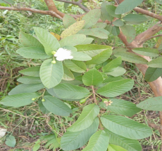
Fig. 1. Guava leaves
Methods
Preparation of the extract
A total of 100 g of powdered guava leaf sample was extracted with 96 % ethanol. The extraction process was subjected to the ultrasonic-assisted extraction method described by Van et al. (2025) [8]. For 100 g of sample, 600 mL of 96 % ethanol was added. Ultrasonic extraction was performed at 50°C for 45 min using an Elma-110 ultrasonic bath (Germany) with a frequency of 37 kHz and ultrasonic power of 150 W. The extraction was repeated three times. After each extraction, the supernatant was filtered through Whatman filter paper, and the residue was used for the next extraction cycle. The combined extracts from the three extractions were concentrated and dried using a rotary vacuum evaporator at 50°C under a pressure of 70 atm to obtain the crude extract coded as GA.100.
Qualitative Phytochemical Screening
To evaluate the presence of bioactive compounds in the GA.US and GA.24 extracts, qualitative chemical tests were carried out according to the methods described by Shaikh and Patil [6] and Tiwari et al. (2011) [9]. The control samples were GA.US and GA.24 extracts diluted to a concentration of 1 mg/mL. The procedures were conducted as follows:
– Polyphenols and tannins test: Fifty microliters of extract solution was mixed with 500 µL of distilled water and 2–3 drops of 5 % FeCl₃ solution. A positive reaction was indicated by the formation of a dark blue precipitate.
– Flavonoids test: Fifty microliters of extract solution was mixed with 500 µL of 10 % Pb(CH₃COO)₂ solution. A yellow precipitate indicated the presence of flavonoids.
– Terpenoids test: Fifty microliters of extract solution was mixed with 500 µL of CH₂Cl₂ and 2–3 drops of concentrated H₂SO₄. A brick-red or green precipitate indicated the presence of terpenoids.
– Quinones test: Fifty microliters of extract solution was mixed with 3–4 drops of 1 M HCl. The appearance of a green precipitate indicated the presence of quinones.
– Coumarins test: Fifty microliters of extract solution was mixed with 750 µL of 10 % NaOH solution. A yellow precipitate indicated the presence of coumarins.
– Saponins test: Fifty microliters of extract solution was mixed with 2 mL of distilled water and a few drops of vegetable oil, then heated at 90°C for 30 min. The formation of a milky emulsion indicated the presence of saponins.
Evaluation of antibacterial activity
The antibacterial activity of the extract was evaluated according to the method described by Tran Chi Linh et al. (2020) [10] against three pathogenic bacterial strains: Bacillus cereus ATCC 11778, Escherichia coli ATCC 11105, and Staphylococcus aureus ATCC 6538. The extract was used at a concentration of 10 %. Agar wells were prepared, and 80 µL of extract solution was added into each well. The inhibition zone diameter was measured in millimeters (mm). Kanamycin at a concentration of 100 µg/mL was used as the positive control.
Results and discussion
Qualitative phytochemical screening of guava leaf extract
The qualitative phytochemical screening of the GA.100 extract revealed the presence of polyphenols, tannins, flavonoids, terpenoids, and coumarins. In contrast, saponins and quinones were not detected. In the tests for polyphenols and tannins, flavonoids, and coumarins, clear color changes were observed during the reactions. The results are presented in Table 1.
Table 1
Qualitative screening results of bioactive compounds in guava leaf extract
|
Compounds |
Polyphenol và tanin |
Flavonoid |
Terpenoid |
Quinone |
Coumarin |
Saponin |
|
GA.100 |
+++ |
+++ |
+ |
- |
+++ |
- |
(Note: (+++), (++): strongly present compounds; (+): present; (−): absent).
Table 2
Antibacterial activity of guava leaf extract
|
Microbial strains |
Diameter of inhibition zone (mm) |
|
Bacillus cereus SH46 |
13 |
|
Escherichia coli VTCC12272 |
15 |
|
Staphylococcus aureus VTCC1227 |
8 |
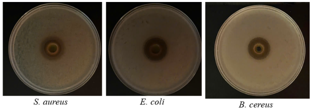
Fig. 2.Antibacterial assay of guava leaf extract
The antibacterial activity of the guava leaf extract, as determined by the agar well diffusion method, is summarized in Table 2 and Fig. 2. The guava leaf extract showed inhibitory effects against all three tested bacterial strains, including Escherichia coli VTCC12272, Bacillus cereus SH46, and Staphylococcus aureus VTCC1227. Among them, the extract exhibited the strongest antibacterial activity against E. coli , with an inhibition zone diameter of 15 mm, followed by B. cereus with an inhibition zone of 13 mm. In contrast, the antibacterial activity against S. aureus was lower, with an inhibition zone diameter of 8 mm. These results indicate that the ethanol extract of guava leaves possesses a relatively broad antibacterial spectrum against both Gram-negative and Gram-positive bacteria. The antibacterial activity may be related to the presence of secondary metabolites such as flavonoids, tannins, polyphenols, and terpenoids detected in the extract. These compounds may alter bacterial cell membrane permeability, inhibit enzyme activity, or cause leakage of intracellular components, thereby reducing bacterial growth.
In this study, E. coli was the most sensitive strain to the guava leaf extract. This result is consistent with the report of Nguyen Thi Bich Thuyen et al., in which guava leaf extract also showed strong inhibitory activity against E. coli [5]. In addition, Le Xuan Duy et al. [4] reported that some compounds isolated from guava leaves, such as guaijaverin and ursolic acid, exhibited strong antibacterial activity against E. coli . For S. aureus , the smaller inhibition zone suggests that this bacterial strain may have higher resistance to the bioactive compounds present in the extract. Differences in bacterial sensitivity may be associated with variations in cell wall structure and physiological characteristics between Gram-positive and Gram-negative bacteria.
Conclusion
In conclusion, the findings of this study demonstrate that the ethanol extract of Dong Du guava leavesserves as a promising natural source of potent antibacterial agents. The presence of diverse phytochemicals, coupled with significant inhibitory activity against both Gram-negative and Gram-positive pathogens, provides a strong scientific foundation for further research. Future studies should focus on the isolation and characterization of specific bioactive compounds to facilitate the development of high-value applications in the food and pharmaceutical industries.
References:
- Kumar, M., Tomar, M., Amarowicz, R., et al. (2021). Guava (Psidium guajava L.) leaves: Nutritional composition, phytochemical profile, and health-promoting bioactivities. Foods, 10(4), 752.
- Nguyen, V. H., & Nguyen, M. C. (2014). Handbook and techniques for caring for guava trees. Agriculture Publishing House. (In Vietnamese).
- Do, T. T. N., Doan, T. K. T., & Le, T. T. (2024). Total polyphenol content, antioxidant, antibacterial and antifungal activities of Psidium guajava L. extract. (In Vietnamese). Can Tho Journal of Science and Technology, 70, 30–38.
- Le, X. D., Nguyen, T. K. A., Le, T. T. L., & Vu, T. T. L. (2025). Chemical constituents and antibacterial activity of compounds isolated from ethyl acetate extract of guava leaves (Psidium guajava L.) collected in Hung Yen province, Vietnam. (In Vietnamese). JNU Journal of Science and Technology, 61(5B), 352–362.
- Nguyen, T. B. T., Cao, L. N. H., Doan, V. H. T., & Tran, T. M. (2023). Study on chemical composition and biological activity of Psidium guajava L. leaf extract. (In Vietnamese). CTU Journal of Innovation and Sustainable Development, 59(Special issue: Engineering and Technology), 129–133.
- Shaikh, J. R., & Patil, M. (2020). Qualitative tests or preliminary phytochemical screening: An overview. International Journal of Chemical Studies, 8(2), 603–608.
- Tiwari, P., Kumar, B., Kaur, M., et al. (2011). Phytochemical screening and extraction: A review. International Pharmaceutical Sciencia, 1, 89–106.
- Van, N. T. C., Hoang, G. B., Nguyen, T. T. T., Pham, T. K. V., Vu, T. P., Tran, V. T., & Cao, T. H. (2025). Nghiên cứu tổng hợp xanh nano bạc từ cao chiết lá vú sữa Chrysophyllum cainito thu hái tại tỉnh Nam Định, Việt Nam. Tạp chí Khoa học và Công nghệ, Trường Đại học Công nghiệp Hà Nội, 61(5B), 294–299.
- Tiwari, P., Kumar, B., Kaur, M., et al. (2011). Phytochemical screening and extraction: A review. International Pharmaceutical Sciencia, 1, 89–106.
- Tran, C. L., Dai, T. X. T., Pham, K. N. H., Vo, T. T. A., Luu, T. D., & Tran, T. M. (2020). Khảo sát hoạt tính sinh học của cao chiết từ rễ cây cỏ sen (Miliusa velutina). In Proceedings of the National Biotechnology Conference 2020 (pp. 1–7). Hue University Publishing House. (In Vietnamese).